

Preventive Eye Health Services at Pinehouse Eyecare
Numerous sight-threatening conditions develop silently without initial pain or noticeable visual loss. Scheduling regular comprehensive eye exams is vital for identifying underlying changes before they permanently compromise your sight.
At Pinehouse Eyecare, our optometry team offers detailed evaluations, long-term monitoring, and customized care plans for various eye diseases. We incorporate modern diagnostic technology to support your comfort and health throughout every visit.
Schedule your comprehensive eye exam today.
Diagnostic Screening Tools for Silent Conditions
Eye diseases frequently advance beneath the surface, causing minimal discomfort or obvious symptoms during their initial stages. Our clinical equipment enables our team to inspect internal eye structures and capture early indicators of disease that might otherwise remain undetected.
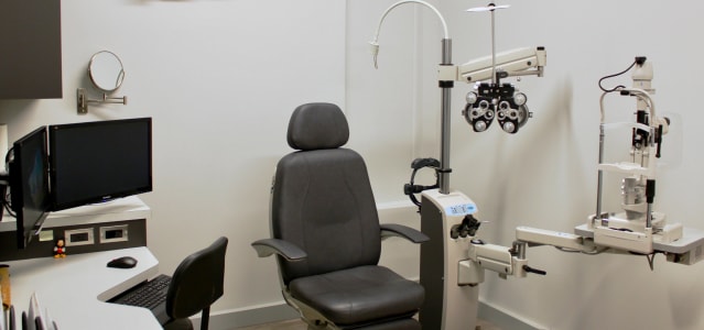
During routine eye exams, our office uses Optomap ultra-widefield imaging, OCT scans, and FDT Matrix visual field evaluations. These diagnostic tools assist in detecting signs of common conditions, such as glaucoma, age-related macular degeneration (AMD), and diabetic retinopathy early enough to begin targeted management.

Glaucoma
Glaucoma refers to a group of progressive eye diseases that harm the optic nerve. This nerve is the primary pathway carrying visual signals from the eye to the brain.
Elevated fluid pressure inside the eye is a common contributing factor to glaucoma, though damage can also occur when intraocular pressure remains within normal limits. Because glaucoma typically erodes peripheral vision first, considerable damage can take place before you notice changes in your central field of view, making it a leading cause of irreversible vision loss.
Open-angle glaucoma is the most frequent variation, progressing gradually as the internal drainage network becomes restricted over time.
Angle-closure glaucoma happens when the iris shifts forward, abruptly blocking fluid drainage and triggering a rapid, severe rise in eye pressure that requires immediate emergency care.
Normal-tension glaucoma occurs when optic nerve fibres suffer damage despite normal fluid pressure readings inside the eye.
Detecting glaucoma relies on a combination of targeted clinical tests to evaluate intraocular pressure and structural nerve integrity.
Tonometry measures internal fluid pressure, while OCT scans generate detailed cross-sectional images of the optic nerve head. Additionally, visual field testing with the FDT Matrix checks your side vision to locate subtle areas of reduced light sensitivity.
Although existing optic nerve damage cannot be restored, ongoing clinical management helps stabilize internal pressure to prevent further sight loss.
Care typically starts with daily prescription eye drops designed to reduce fluid production or promote better drainage. In certain situations, your optometrist may recommend oral medications, laser procedures, or surgical referrals to support long-term pressure control.
Cataracts
A cataract is the gradual clouding of the eye’s natural crystalline lens, situated directly behind the iris and pupil. As proteins inside the lens clump together with age, light cannot pass cleanly to the retina, causing objects to look hazy or out of focus.
Cataracts progressively reduce visual sharpness, dull colour contrast, and create distracting glare, particularly during nighttime driving.
Common indicators of cataract development include:
- Blurry, dim, or double vision
- Faded, yellowed, or washed-out colour perception
- Increased sensitivity to bright sun or indoor lighting
- Halos or ring reflections around light sources
- Difficulty seeing clearly in low-light environments
Natural aging is the leading factor in cataract development as lens proteins break down over time.
Prolonged exposure to ultraviolet (UV) sunlight, diabetes, tobacco use, and excessive alcohol consumption also accelerate lens clouding.
Additionally, long-term corticosteroid use or past ocular injuries can heighten your risk of developing cataracts earlier in life.
In early stages, cataract symptoms can often be managed by updating your eyeglass prescription and incorporating brighter lighting for reading or close work.
When lens clouding progresses to the point of interfering with daily routines, cataract surgery offers an effective solution. An ophthalmologist replaces the cloudy lens with a clear artificial intraocular lens, and our clinic provides comprehensive pre-operative guidance and post-operative follow-up care.
Age-Related Macular Degeneration (AMD)
Age-related macular degeneration (AMD) impacts the macula, the specialized central region of the retina responsible for clear, straight-ahead vision. As AMD advances, it hinders daily tasks like reading fine print, recognizing faces, driving, or performing detailed work.
Individuals with AMD often notice that central vision becomes distorted, straight lines appear bent or wavy, or dark spots appear in their field of view.
Dry AMD is the most common form, characterized by slow thinning of macular tissue and the accumulation of yellow metabolic deposits called drusen.
Wet AMD is a less common but more aggressive form where fragile, abnormal blood vessels grow underneath the retina and leak fluid or blood into the macula, causing rapid central vision changes that demand immediate medical attention.
Age is the primary risk factor for macular degeneration, with incidence rising significantly in adults over 50.
Family history and genetic factors play a strong role, while smoking dramatically increases your likelihood of developing the condition.
Uncontrolled hypertension, elevated cholesterol, and poor dietary habits also contribute to macular damage over time.
Evaluations for AMD take place during comprehensive eye exams using high-resolution retinal imaging and OCT scans to check macular layers for drusen or fluid accumulation.
An Amsler grid test is also used to identify distorted grid lines or blind spots in your central visual field. These diagnostic tools help us detect subtle structural changes and track macular health over time.
Treatment protocols depend on whether you are diagnosed with dry or wet AMD.
For dry AMD, targeted nutritional supplements based on the AREDS2 formulation and specialized therapies like MacuMira help support retinal cell health and help preserve central sight.
For wet AMD, prompt referral for anti-VEGF injections or laser treatments helps seal leaking vessels and control macular swelling.
Flashes and Floaters
Flashes and floaters are visual symptoms associated with changes inside the vitreous, the clear gel-like fluid filling the inside of your eye.
While occasional floaters are common as the vitreous ages, a sudden onset or drastic shift in these visual symptoms can indicate an underlying retinal concern that requires prompt clinical attention.
Flashes present as brief streaks, arcs, or sparks of light across your visual field, resembling lightning or camera flashes. They occur when the shrinking vitreous gel pulls or tugs against the light-sensitive retina. While occasional flashes can be benign, a sudden increase in frequency suggests retinal traction that must be evaluated promptly.
Floaters appear as small specks, squiggles, web-like strands, or dark spots drifting slowly across your line of sight. They are caused by microscopic protein strands within the vitreous gel casting small shadows onto the retina. Most floaters are harmless signs of aging, but a sudden burst of new floaters can signal a retinal tear.
Seek urgent eye care if you experience a sudden wave of new floaters, frequent light flashes, or a dark curtain-like shadow obscuring your side vision. These warning signs can indicate a retinal tear or detachment, which requires immediate treatment to prevent permanent sight loss. If you notice these symptoms, contact our clinic right away for an emergency evaluation.
Conjunctivitis
Conjunctivitis, commonly called pink eye, involves inflammation or infection of the conjunctiva—the clear membrane lining your eyelid and covering the white part of your eye.
It causes small blood vessels in the tissue to dilate, leading to redness, irritation, and discharge. Because conjunctivitis can arise from several distinct causes, a proper clinical examination is necessary to guide effective treatment.
Allergic conjunctivitis develops when your eyes react to airborne allergens like pollen, pet dander, dust, or mold spores. It usually affects both eyes at once, producing intense itching, watery discharge, redness, and swollen eyelids without being contagious.
Management includes allergen avoidance, cool compresses, and lubricating or over-the-counter antihistamine eye drops.
Viral conjunctivitis is highly contagious and frequently accompanies common respiratory infections or colds. Symptoms typically include watery discharge, redness, a gritty sensation, and light sensitivity, often beginning in one eye before spreading to the other.
Since antibiotics cannot treat viral infections, management focuses on comforting therapies, cold compresses, artificial tears, and strict hygiene to prevent transmission.
Bacterial conjunctivitis stems from bacterial infection and is marked by thick, yellow or greenish discharge that can cause the eyelids to stick shut overnight. It can develop in one or both eyes and spreads easily through contact with contaminated hands or personal items.
Treatment involves prescription antibiotic eye drops or ointments to resolve the infection and relieve symptoms quickly.
Schedule Your Next Eye Assessment
Eye diseases can impact visual clarity and long-term sight, but many can be managed effectively when detected early. From glaucoma and cataracts to macular degeneration, consistent clinical evaluations offer a strong defence against progressive vision loss.
We recommend scheduling a comprehensive eye exam every 1 to 2 years, or as directed by your optometrist, to maintain proactive care for your eyes.
Frequently Asked Questions About Eye Disease
We recommend scheduling a comprehensive eye exam every 1 to 2 years to check for early signs of eye disease and monitor changes in your sight. If you have a family history of eye conditions or notice new visual symptoms, you should book an appointment at Pinehouse Eyecare regularly.
Yes, many serious eye conditions like glaucoma, macular degeneration, and diabetic retinopathy can progress silently without pain or early vision loss. Routine comprehensive eye exams every 1 to 2 years allow your optometrist to spot subtle structural changes early and manage them before permanent damage occurs.
Warning signs of eye disease include persistent blurriness, dark spots in your central vision, sudden flashes of light, new floaters, or loss of peripheral vision. If you notice any of these warning signs, please book a comprehensive eye exam with our team right away.
In many cases, vision loss caused by conditions like glaucoma or advanced macular degeneration is permanent and cannot be reversed. However, early detection through regular eye exams allows your optometrist to implement management strategies that help preserve your remaining sight.
Having a family history of conditions such as glaucoma, age-related macular degeneration, or cataracts significantly increases your likelihood of developing similar issues. If eye disease runs in your family, schedule a comprehensive exam at Pinehouse Eyecare to establish an appropriate baseline and monitoring routine.
Optometrists identify eye conditions using advanced diagnostic tools, including digital retinal imaging, OCT scans, visual field testing, and dilated eye evaluations. Booking a comprehensive eye exam every 1 to 2 years gives our optometry team the opportunity to assess your complete eye health in detail.

Visit Our Location
We’re located on the corner of Pinehouse Drive and Primrose Drive. Find us just across the street from Umea Vast Park. Free parking is available in front of the clinic.
Our Address
Contact Information
- Phone: 306-242-4186
- Fax: 306-242-5586
- Email: pecadmin@yxevision.ca
After Hours Emergencies: 306-371-9911
Hours of Operation
Take a Glance
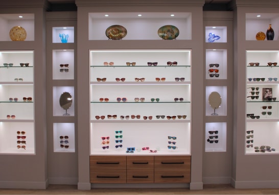
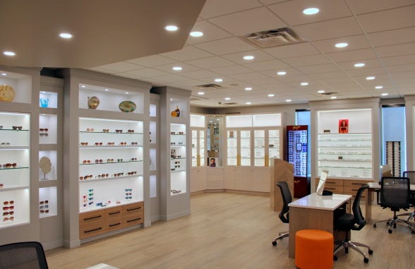
Inside Pinehouse Eyecare


See What Our Patients Are Saying
